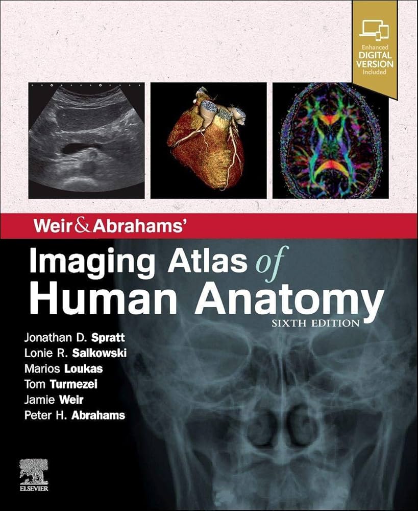
Weir & Abrahams’ Imaging Atlas of Human Anatomy is a vital resource for ST1 radiology registrars preparing for the FRCR Part 1 Anatomy exam as it provides a comprehensive inventory of annotated anatomy on different modalities of medical imaging (plain radiographs, CT, MR, ultrasound etc.) covering the entire human body. It is also useful for medical students and other healthcare professionals seeking a clear and comprehensive understanding of human anatomy through medical imaging. The book, now in its sixth edition, has established itself as one of the most detailed and user-friendly atlases for correlating clinical anatomy with radiological images.
Overview of Content
Weir & Abrahams’ Imaging Atlas of Human Anatomy offers an in-depth exploration of human anatomy by combining high-quality imaging from plain radiographs, CT, MRI, and ultrasound with detailed anatomical illustrations. It covers every part of the body, breaking down complex anatomical regions into manageable sections with accompanying imaging studies that illustrate both normal and pathological structures.
Each chapter is arranged by body region, making it easy for readers to focus on specific areas of interest, whether it’s the head and neck, thorax, abdomen, or musculoskeletal system.
Key Features
- Comprehensive Imaging Collection: One of the most striking features of Weir & Abrahams’ Imaging Atlas of Human Anatomy is its extensive array of images. It includes thousands of radiological images taken using various modalities such as plain radiographs, CT, MRI, and ultrasound, offering readers a real-life perspective of how anatomy appears in clinical practice. The images are carefully selected and annotated, ensuring that readers can quickly understand the anatomical structures depicted.
- Correlated Imaging and Anatomy: The hallmark of this atlas is its ability to correlate medical imaging with detailed anatomical descriptions. Each image is paired with clear, precise anatomical illustrations, making it easy to compare the radiological findings with theoretical anatomical knowledge. This is particularly useful for understanding the spatial relationships between different organs and tissues in the body.
- High-Quality Annotations: Every image is accompanied by detailed and well-labelled annotations, which guide the reader through the complexities of human anatomy. These annotations make it easier to identify structures and understand their clinical relevance. They are presented in a clear, colour-coded manner, making the atlas highly accessible.
- Clinical Relevance: The atlas bridges the gap between basic anatomical knowledge and its application in clinical practice. The authors frequently include information on how anatomical structures appear in pathological states, which helps clinicians to recognise disease processes on imaging. This feature enhances the practical value of the book for both students and professionals.
Pros of the Book
- Detailed and Accurate Imaging: The wealth of high-resolution images in Weir & Abrahams’ Imaging Atlas of Human Anatomy is one of its strongest points. The images, drawn from real-life clinical cases, offer a realistic and accurate portrayal of human anatomy, giving readers a true sense of how anatomy is visualised in clinical practice.
- User-Friendly Layout: The book’s structure is particularly appealing for those using it as a reference guide. Its organisation by anatomical region, combined with clear headings and subheadings, allows users to navigate easily through the content. This is particularly useful for medical students and clinicians in need of quick information.
- Correlative Approach: The inclusion of both radiological images and anatomical illustrations helps bridge the gap between theoretical learning and practical application. This correlative approach is especially helpful for students and professionals who need to compare their anatomical knowledge with what they encounter on imaging studies.
- Comprehensive Coverage: From the head to the toes, this atlas covers all areas of the human body. It doesn’t just stop at the basics but goes deeper into more complex regions such as the neurovascular structures, which are often difficult to interpret in imaging. This makes the book suitable for both beginners and more advanced learners.
Cons of the Book
- High Price Point: As with many comprehensive medical texts, Weir & Abrahams’ Imaging Atlas of Human Anatomy comes with a significant price tag. This could be a drawback for new ST1 radiology registrars on a budget. However, considering the wealth of information and high-quality imaging provided, it can be seen as a worthwhile investment for serious learners.
- Limited to Imaging: Although the book excels at presenting anatomy through radiological images, it is primarily focused on that modality. Those looking for a more traditional, detailed anatomical textbook with extensive cadaveric dissections or histological details may find this resource lacking in those areas.
- Not as In-Depth on Pathologies: While the book provides basic information on how pathological changes appear on imaging, it does not delve deeply into disease processes or their mechanisms. This may leave some readers wanting more information on the clinical implications of certain findings.
- Less Suitable for Beginners in Anatomy: For absolute beginners in anatomy, particularly those without any experience with imaging, the amount of information in this atlas could be overwhelming. The book assumes a certain level of anatomical knowledge and might be more useful as a supplementary tool rather than a primary text for those just starting out.
Conclusion
Weir & Abrahams’ Imaging Atlas of Human Anatomy is a comprehensive, well-structured, and visually stunning resource for learning and teaching human anatomy through imaging. Its wealth of high-quality images, combined with clear and concise anatomical descriptions, makes it an indispensable tool for ST1 radiology registrars preparing to sit the FRCR Part 1 Anatomy examination.
While the book is highly specialised and may not be suitable for those looking for a broader anatomical text, its focus on practical application and clinical relevance makes it a standout resource in the field. Despite its high price, the quality and depth of information provided make it a worthy investment for anyone serious about mastering human anatomy through imaging.
Please note: Our website is funded by user engagement. As such, we may receive a small commission when you purchase a qualifying product via an affiliate link.
