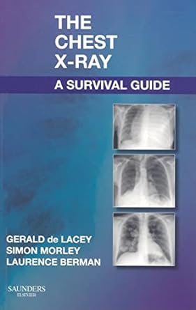
The Chest X-Ray: A Survival Guide is one of our recommended textbooks for new radiology registrars, looking to develop their understanding and interpretation of chest X-rays. It is a concise read which covers the common chest X-ray findings.
Overview of Content
The book is divided into easily digestible sections, starting with the basics of reading chest X-rays, which includes a review of normal anatomy and how it appears on an X-ray. It then progresses into more advanced concepts, such as recognising abnormal patterns, diagnosing pathologies, and understanding differential diagnoses.
What sets The Chest X-Ray: A Survival Guide apart from similar texts is its strong clinical focus. Rather than merely presenting the theory of radiographic imaging, the authors use their extensive experience in radiology to provide practical tips and insights. They include numerous case studies and real-life examples to illustrate common and rare findings on chest X-rays. The inclusion of high-quality images, along with clear and concise explanations, helps solidify the reader’s knowledge and confidence in interpreting X-ray results.
Key Features
- Systematic Approach: One of the major strengths of The Chest X-Ray: A Survival Guide is its structured methodology in teaching chest X-ray interpretation. The step-by-step approach ensures that readers develop a strong foundational understanding before moving on to more complex cases. Each chapter is designed to build on the previous one, making the learning curve smooth and manageable.
- Case Studies: Real-world case studies feature heavily in this guide, offering practical examples of how to apply the knowledge gained. These case studies are incredibly useful for developing diagnostic skills and learning how to identify various conditions ranging from lung infections to pneumothorax and heart failure.
- Annotated Images: A standout feature is the extensive use of annotated chest X-rays. These annotations help highlight key areas of interest, making it easier for readers to follow along with the explanations and apply their learning in practice.
- Differential Diagnosis Lists: The book includes handy checklists and differential diagnosis lists, which provide a quick reference point during interpretation. This is particularly useful for medical professionals who are in the early stages of developing their diagnostic acumen.
- Concise Explanations: Despite its detailed content, the book maintains a concise and easy-to-understand writing style. Medical jargon is explained in straightforward terms, making it suitable for a wide audience, from beginners to experienced clinicians looking to refresh their knowledge.
Pros of the Book
- Practical Focus: The book’s emphasis on practical applications and real-life scenarios makes it highly relevant to clinical practice. This is not a purely academic text but one that actively encourages the reader to think like a practising radiologist.
- Ease of Use: For beginners, the logical progression from basic anatomy to more advanced pathology makes the book easy to follow. Even complex concepts are broken down into manageable chunks, which can be digested at the reader’s own pace.
- High-Quality Images: The clear, high-resolution X-ray images, complete with annotations, are one of the book’s biggest strengths. These visuals help reinforce the text and provide a reference for real-life X-ray interpretation.
- Checklists and Mnemonics: The inclusion of checklists and mnemonics is a useful tool. These concise memory aids help consolidate learning and provide a quick reference guide.
- Compact and Portable: Despite covering a wide range of topics, the book is relatively compact, making it a handy reference guide to carry in a clinical setting. It’s also a good size for dipping into between clinical shifts or while on the go.
Cons of the Book
- Limited Depth on Some Topics: While the book covers a broad range of conditions, some readers may find the depth on rarer conditions or advanced techniques somewhat limited. This book certainly does not have enough content on CXRs for a radiologist and that is why it should be used as an introductory text for new registrats.
- Lack of Online Resources: In an age where supplementary online content is often provided alongside textbooks, the absence of any associated digital learning tools or online resources is notable. Interactive platforms, video tutorials, or quizzes could have enhanced the learning experience.
- Outdated in Certain Areas: Published in 2008, the book may feel slightly dated in some areas, particularly as medical imaging technology has advanced significantly over the years. While the principles of chest X-ray interpretation remain largely unchanged, newer editions incorporating the latest imaging techniques, such as CT imaging, would be beneficial.
- Geared Towards Beginners: The book is more suited to those just starting their journey in radiology or those looking to brush up on the basics.
Who Would Benefit from This Book?
The Chest X-Ray: A Survival Guide is ideal for new radiology trainees, as well as other doctors and medical students. It serves as a strong foundational text for those beginning their radiology training or those looking to improve their confidence in reading chest X-rays.
Conclusion
In summary, The Chest X-Ray: A Survival Guide is a valuable tool for new radiology trainees. Its practical approach, user-friendly structure, and emphasis on real-world applications make it an accessible and useful reference guide. While it may lack depth in some areas and feels slightly dated in light of modern advancements in radiology, it remains a solid and reliable resource for learning the basics of chest X-ray interpretation.
Please note: Our website is funded by user engagement. As such, we may receive a small commission when you purchase a qualifying product via an affiliate link.
